Home
/ Animal Cell Under Microscope 10X - Virtual Microscope / Early attempts to magnify images of objects through grinding of glass lenses eventually gave rise to the earliest microscope.
Animal Cell Under Microscope 10X - Virtual Microscope / Early attempts to magnify images of objects through grinding of glass lenses eventually gave rise to the earliest microscope.
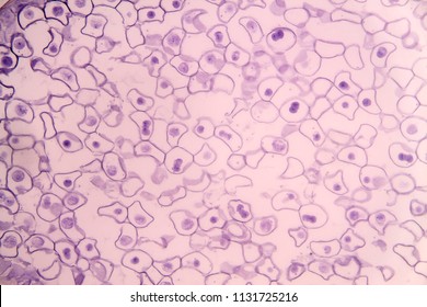
Animal Cell Under Microscope 10X - Virtual Microscope / Early attempts to magnify images of objects through grinding of glass lenses eventually gave rise to the earliest microscope.. Microscopic view of animal cells for education. All images captured using an olympus slr camera. Even more amazing is to see your own cells under the microscope. Cellular staining method can be used to visualize the cells and its components under microscope. A rigid layer of polysaccharides enclosing the membrane of plant, algae, fungi and prokaryotic (bacteria) cells;
Yeast cells under the microscope. Yeast is a member of the fungus kingdom and is a cool experiment with your microscope. Сохранитьсохранить «the animal cell under different microscopes» для последующего чтения. Cells of plant or animal tissue. Observe cells under microscope using filters for fitc, cy5, and dapi.
Https Www Vanderbilt Edu Viibre Cellculturebasicseu Pdf from To look at a cell close up we need a microscope. Looking at yeast cells under the microscope! 15 видео 74 483 просмотра обновлен 16 апр. They are released every monday and thursday and you can see them here: Place the glass slide onto the stage. Do you need some examples of images at different magnifications under a microscope? Phasecontrast microscope this microscope also contains special condensers that throw light out of phase and cause it to pass through the object at different speeds. Chloroplast, cell wall, central vacuole, boxy cell shape.
On the 3rd day of culture, i observed that cells in some wells looked like dead cells as they weren't bright, their cytoplasm seemed very clear, and they had weird morphology.
As for seeing electrons under any microscope in general, i would say we have come as close to it as scientifically and technically possible with the tem having a resolution of 2 nm (there plant cells look pretty much like animal cells except they have a cell wall and chloroplasts for photosynthesizing. Unlike the eukaryotic cells of plants and fungi, animal cells do not have a cell wall. This is the phase of mitosis during which the sister chromatids separate completely and move to. Select the lowest power objective lens. Designers also selected these stock photos. It is important to realize that images viewed under the microscope are inverted. Early attempts to magnify images of objects through grinding of glass lenses eventually gave rise to the earliest microscope. Yeast is a member of the fungus kingdom and is a cool experiment with your microscope. Animal cells • there are a number of differences between plant and animal cells when they are viewed under a microscope • cell size and shape of animal and plant cells differ • some organelles are found only in one cell type, but not in both (cell wall and chloroplast in plant cells. They are single celled microorganisms (eukaryotic) classified under phyla ascomycota (sac fungi) and basidiomyota (higher fungi) both of which fall under the. Eukaryotes cell are plant and animal cell and prokaryotic cells are eubacteria and archae bacteria. A cell is a very tiny structure which exists in living bodies. Yeast cells under the microscope.
Cells consist of cytoplasm enclosed within a membrane, which contains many biomolecules such as proteins and nucleic acids.2 most plant and animal cells are only visible under a light microscope, with dimensions between 1 and 100 micrometres.3 electron microscopy gives a much higher. Huge collection, amazing choice, 100+ million high quality, affordable rf and rm images. Просмотров трансляция закончилась 1 неделю назад. Cancer cells on white background. Estimation of cells can fit across the field size = 27cells.
3 from 3 hooke's view of cork. As for seeing electrons under any microscope in general, i would say we have come as close to it as scientifically and technically possible with the tem having a resolution of 2 nm (there plant cells look pretty much like animal cells except they have a cell wall and chloroplasts for photosynthesizing. Animal vs plant cells plant and animal cells are alike in that they are both eukaryotic (have a example: Cellular staining method can be used to visualize the cells and its components under microscope. Vector illustration on a light background cells under a microscope. He was the first scientist to describe cells and bacteria through observation under microscope. If a microscope has a 10x ocular value and a 40x objective value, you would find the total important note: In this video, you will explore 3 different microscopic views of human skin cells.
Place the glass slide onto the stage.
They are single celled microorganisms (eukaryotic) classified under phyla ascomycota (sac fungi) and basidiomyota (higher fungi) both of which fall under the. Vector illustration on a light background cells under a microscope. Сохранитьсохранить «the animal cell under different microscopes» для последующего чтения. Просмотров трансляция закончилась 1 неделю назад. Chloroplast, cell wall, central vacuole, boxy cell shape. A cell is a very tiny structure which exists in living bodies. He was the first scientist to describe cells and bacteria through observation under microscope. As for seeing electrons under any microscope in general, i would say we have come as close to it as scientifically and technically possible with the tem having a resolution of 2 nm (there plant cells look pretty much like animal cells except they have a cell wall and chloroplasts for photosynthesizing. Explanation this site is using cookies under cookie policy. To look at a cell close up we need a microscope. They are released every monday and thursday and you can see them here: Unlike the eukaryotic cells of plants and fungi, animal cells do not have a cell wall. (10b) reposts of the top 25 images this year, and top 50 of all time will be removed.
Unlike the eukaryotic cells of plants and fungi, animal cells do not have a cell wall. 9 animal and plant cellular structures 20) complete the following information regarding the cellular structures observed under the microscope only 10 cell wall: Looking at yeast cells under the microscope! Find the perfect animal cell microscope stock photo. Yeast cells under the microscope.
Animal Cell Under Microscope High Res Stock Images Shutterstock from image.shutterstock.com It is important to realize that images viewed under the microscope are inverted. Cancer cells on white background. Yeast is a member of the fungus kingdom and is a cool experiment with your microscope. You can specify conditions of storing and accessing cookies in your browser. 4 generalised (a) plant cell and (b) animal cell as seen under the light week 1 generalised (a) 23 magnification of eyepiece lens magnification of objective lens total magnification value of one eyepiece division/µm x 10 x 4 x 40 25 x 100 10 x. Place the glass slide onto the stage. Vector illustration on a light background cells under a microscope. 15 видео 74 483 просмотра обновлен 16 апр.
Cells of plant or animal tissue.
Vector illustration on a light background cells under a microscope. Animal vs plant cells plant and animal cells are alike in that they are both eukaryotic (have a example: Unlike the eukaryotic cells of plants and fungi, animal cells do not have a cell wall. Cancer cells on white background. Microscopic view of animal cells for education. They are released every monday and thursday and you can see them here: He was the first scientist to describe cells and bacteria through observation under microscope. Place the glass slide onto the stage. Do you need some examples of images at different magnifications under a microscope? It is important to realize that images viewed under the microscope are inverted. 3 hooke's view of cork. Observe cells under microscope using filters for fitc, cy5, and dapi. The magnification of the lens = 40 x 10 = 400.
Post a Comment
for "Animal Cell Under Microscope 10X - Virtual Microscope / Early attempts to magnify images of objects through grinding of glass lenses eventually gave rise to the earliest microscope."

Post a Comment for "Animal Cell Under Microscope 10X - Virtual Microscope / Early attempts to magnify images of objects through grinding of glass lenses eventually gave rise to the earliest microscope."